Cement (PMMA) Extrusion into Femoral Nutrient Vessels during Total Hip Arthroplasty: Case Series
Hemanth Kumar Venkatesh and Mohammad Shoaib
DOI10.4172/2469-6684.100032
Hemanth Kumar Venkatesh* and Mohammad Shoaib
Consultant Orthopaedic Surgeon, Basildon and Thurrock University Hospital, Nethermayne, Basildon, UK
- *Corresponding Author:
- Hemanth Kumar Venkatesh
Consultant Orthopaedic Surgeon, Basildon and Thurrock University Hospital
Nethermayne, Basildon SS16, UK
Tel: +441268524900/2748
Fax: +441268394654
E-mail: vhemanthkumar09@gmail.com
Received date: October 15, 2016; Accepted date: November 09, 2016; Published date: November 11, 2016
Citation: Venkatesh HK, Shoaib M. Cement (PMMA) Extrusion into Femoral Nutrient Vessels during Total Hip Arthroplasty: Case Series. J Bone Rep Recommendations. 2016, 2:4.Doi: 10.4172/2469-6684.100032
Copyright: © 2016 Venkatesh HK, et al. This is an open-access article distributed under the terms of the Creative Commons Attribution License, which permits unrestricted use, distribution, and reproduction in any medium, provided the original author and source are credited.
Keywords
Hip arthroplasty; Hip replacement; Proximal femur; Osteoarthritis; Polymerisation; Radiograph
Abbreviations
THA: Total Hip Arthroplasty; PMMA: Polymethylmethacrylate; CT: Computerized Tomography
Introduction
Current concepts in cement Total Hip Arthroplasty advocates optimization of mechanical interlock of cement (PMMA) with trabecular bone. The Third generation cementing technique has been developed to achieve this with the use of cement gun retrograde cementing within the proximal femur and pressurization of the cement with wedge or surgeon’s thumb. The forces generated by cement compression during total hip replacement reach peak pressures that varied between 122 kPa and more than 1500 kPa. The high intramedullary pressures encountered can lead to extrusion of cement through the nutrient foramina in the femoral cortex and into the nutrient vessels, resulting in the retrograde arteriovenogram. This has been reported in the literature as a rare phenomenon. Published case reports [1,3-9] have highlighted the extrusion of cement into the nutrient vessels either as intravenous extrusion due to the appearance of valvular constriction or as extravascular extrusions of PMMA through a femoral vascular channel.
We report two cases of extrusion of PMMA into the nutrient vessels of the femur following uncomplicated cemented total hip arthroplasty. The radiographic appearance corresponds to the vascular supply of the proximal femur. The cases have been followed up for more than 6 months with no adverse effects.
Case Reports
Case 1
A 73 year old female presenting with right hip osteoarthritis underwent cemented Exeter (Stryker US) total hip arthroplasty through a posterior approach. An Exeter contemporary cup (Stryker, US) utilized along with the Exeter V40, 37.5 sizes 0 (Stryker US) femoral components. The femoral canal prepared, cleaned with pulse lavage, dried and a size 12 mm cement restrictor inserted (cement plug Stryker) two centimeters distal to the tip of the femoral component. It was followed by retrograde introduction of standard viscosity cement (Refobacin, Biomet) into the femoral cavity using 3rd generation cementing technique at 2 min of polymerization. The pressurization was performed with the wedge adapter of the cement gun (Stryker UK cement gun). The femoral component introduced at 3-4 min. No untoward intraoperative events were noted. The patient made a good recovery from the surgery.
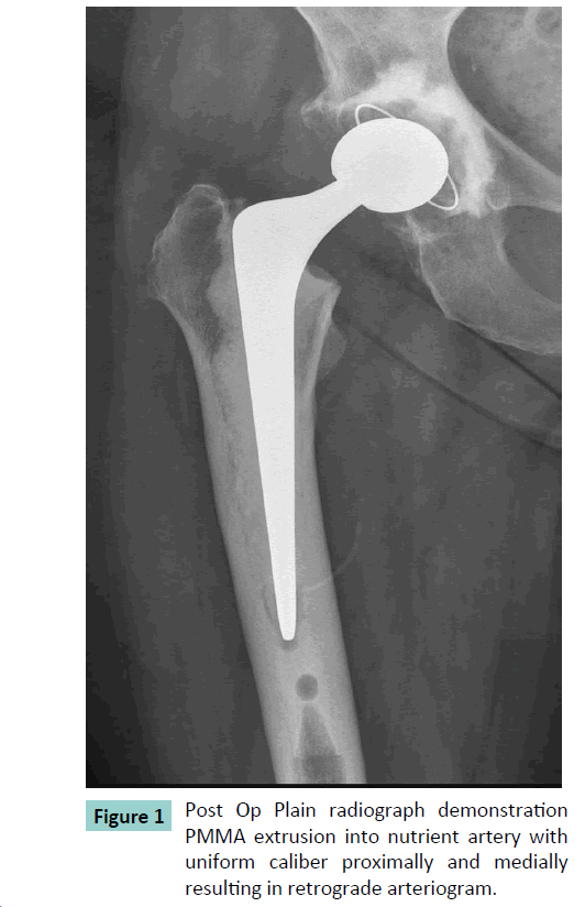
The Post-operative radiograph showed a linear radio opacity communicating with the posterior aspect of the femoral shaft. This extended proximally and medially for approximately about 5cm and was of uniform caliber suggesting PMMA within a blood vessel. There were no valvular constriction evident, indicating that the pressurized cement had produced a retrograde arteriogram of the nutrient artery of the femur (Figure 1).
Case 2
A 79 year old female presenting with bilateral hip osteoarthritis underwent left hip cemented Exeter (Stryker US,) total hip arthroplasty through a posterior approach. An Exeter contemporary cup (Stryker, US) utilized along with the Exeter V40, 37.5 sizes 0 (Stryker US) femoral components. The femoral canal was prepared similar to the previous case with 3rd generation, cement technique and a size 10 cement restrictor [cement plug, Stryker] was utilized. The standard viscosity cement (Refobacin, Biomet) introduced into the femoral canal at 2 min of polymerization. The pressurization was performed with the wedge adapter of the cement gun (Stryker UK cement gun). The femoral component was introduced at 3 min of polymerization. No untoward intraoperative events were noted. The patient made a good recovery from the surgery.
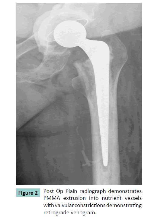
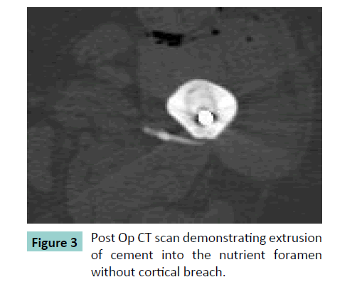
The Post-operative radiograph showed a linear radio opacity communicating with the posterior aspect of the femoral shaft. This extended proximally and medially for approximately about 8cm and varying caliber suggesting PMMA within a blood vessel. There were valvular constrictions noted along the course indicating that the pressurized cement had produced a retrograde venogram of the nutrient vessels of the femur. ACT scan was performed on second post-operative day and showed extrusion of PMMA along the course of nutrient vessel of the femur and no breach in the distal cortex ruling out per prosthetic fracture (Figures 2 and 3).
Discussion
Schiessel and Zweymuller [5] reported the presence of nutrient canal vessel in 26.4% radiographs of preoperative and postoperative radiographs of hip replacement and also reported its anatomical location about 170 ± 25 mm distal to the greater trochanter. The cadaveric study performed by Faruk et al. [6] in his studies of 20 fresh human cadavers reported the nutrient artery and its foramen arising in 166 ± 10 mm from the greater trochanter. This was a branch of second perforating artery from deep femoral artery. The femur was divided into the sixths and the position of the nutrient artery and foramen was found to lie consistently in the third sixth.
Nagel et al. [7] in their anatomical study noted that the nutrient artery foramen contains 1 artery and 2 veins. The extrusion of PMMA at the nutrient artery foramen [8-11] has been described by many authors with plain radiographs. All the case was reported in female patients.
Knight et al. [10] reported the incidence of cement extrusion is 0.9-1.6% of primary cemented femoral component THA and concluded that they occur in female patients with small stature with small endosteal canal. Our case reports support these finding as both our female patients were small with small endosteal diameter.
Panousis et al. [7] reported PMMA within a small diameter vessel with no valvular constrictions resulting in retrograde arteriogram of the nutrient artery of the femur which coupled with the extent of intraoperative endosteal bleeding. The plain radiographs of the Case 1 in our series confirmed linear extrusion of PMMA along the course of nutrient artery proximally and medially with no valvular constrictions resulting in the retrograde arteriogram of the nutrient artery of the femur. In contrast, we did not encounter any intraoperative endosteal bleeding.
The other authors [1,5,7,8,12,13] have reported intravasated standard viscosity cement characteristics of venous filling in their series of 4 patients, with a common feature of appearance of valves characterized by localized constrictions in the caliber of cement radio opacity. It is well established that the nutrient artery has an accompanying vein or veins, which have been implicated in these series. In our case 2, the plain radiograph demonstrated the extrusion of PMMA along the nutrient vessels with the appearance of valvular constriction and CT scan confirmed the cement intravasation within the nutrient vessel. This appearance is of retrograde venogram of the nutrient vessel as reported. Since the periprosthetic fracture was ruled out by CT scan, an arteriogram was not performed to confirm this as it would have not been benefit to the patient.
Skyrme et al. [12] reported that cement outlines the vessel within whereas cortical defect would result in irregular collection of cement that lies adjacent to bone. Knight et al. [3] reported posterior distal cement extrusion in 8 patients with an incidence of 0.9% to 1.6% of primary cemented femoral component THA. The author noted that the size of the extrusion was greater than the diameter of normal vessels of the area and suggested that extrusion was either extravascular or that the initially intravascular PMMA ruptures the vessel and escapes in the surrounding tissues.
In our case series the cement was intravasated within the nutrient vessels without any irregular collection ruling out the periprosthetic fractures.
Cement extrusion unlikely to affect the long term survival of prosthesis as we can infer from the radiographic appearance that cement has been well pressurized there by optimizing mechanical interlock with the endosteal bone. Furthermore, due to extensive anastomoses of the perforating branches of deep femoral artery, which supply overlying muscles and periosteum segmentally obliteration of the nutrient artery alone is unlikely to lead the problem of vascularity [13]. There has been not no study reported any increased comorbidity due to cement extrusion nor been any reported ischemia due to this phenomenon. Both our patients have recovered well with no complication post-surgery
In summary, cement extrusion into the nutrient foramen is an important differential to be considered in presence of posterior medial cement in the diaphysis of the femur following total hip replacement. This is to differentiate from extra osseous extrusions due to iatrogenic breach of the femoral cortex, which may well have implications regarding either periprosthetic fracture or the long term survival of prosthesis.
References
- Horne JG, Bruce W, Devane PA, Teoh HH (2002) The effect of different cement insertion techniques on the bone-cement interface. J Arthroplasty 17: 579-583.
- Bisignani G, Bisignani M, Pasquale GS, Greco F (2008) Intraoperative embolism and hip arthroplasty: intraoperative transesophageal echocardiographic study. J Cardiovasc Med 9:277-281.
- Elmaraghy AW, Humeniuk B, Anderson GI, Schemitsch EH, Richards RR (1998) The role of methylmethacrylate monomer in the formation and haemodynamic outcome of pulmonary fat emboli. J Bone Joint Surg Br 80:156-161.
- Hiesel C,Norman T, Rupp R (2003) In vitro performance of intramedullary cement restriction in total hip arthroplasty.J Biomech.
- Schiessel A, Zweymuller K (2004) The nutrient artery canal of the femur: a radiological study in patients with primary total hip replacement. Skeletal Radiol 33: 142-149.
- Farouk O, Krettek C, Miclau T, Schandelmaier P, Tscherne H (1999) The topography of the perforating vessels of the deep femoral artery. Clin Orthop Relat Res 368: 255-259.
- Nagel A (1993) The clinical significance of the nutrient artery. Orthop Rev 22: 557-561.
- Panousis K, Young KA, Grigoris P (2006) Polymethylmethacrylate arteriography-A complication of total hip arthroplasty. Acta Orthop Belg 72: 226-228.
- Nogler M, Fischer M, Freund M, Mayr E, Bach C, et al. (2002) Retrograde injection of a nutrient vein with cement in cemented total hip arthroplasty. J Arthroplasty 17: 505-506.
- Knight JL, Coglon T, Hagen C, Clark J (1999) Posterior distal cement extrusion during primary total hip arthroplasty: a cause for concern? J Arthroplasty 14: 832-839.
- Brandser E, el-Khoury G, Riley M, Callaghan J (1995) Intravenous methylmethacrylate following cemented total hip arthroplasty. Skeletal Radiol 24: 493-494.
- Skyrme AD, Jeer PJ, Berry J, Lewis SG, Compson JP (2001) Intravenous polymethyl methacrylate after cemented hemiarthroplasty of the hip. J Arthroplasty 16: 521-523.
- Wang L,Gardner AW,Kweek EBK, Rajamoney GN (2012) Retrograde cement arteriovenogram of nutrient vessels following hemiarthroplasty of the Hip. Acta Orthop Belg 78: 431-435.
Open Access Journals
- Aquaculture & Veterinary Science
- Chemistry & Chemical Sciences
- Clinical Sciences
- Engineering
- General Science
- Genetics & Molecular Biology
- Health Care & Nursing
- Immunology & Microbiology
- Materials Science
- Mathematics & Physics
- Medical Sciences
- Neurology & Psychiatry
- Oncology & Cancer Science
- Pharmaceutical Sciences


